24/7 Tailored Support
Expert assistance. Anytime. Anywhere.

Expert assistance. Anytime. Anywhere.

24/7 ordering with stable, efficient logistics.

Certified products, high-performance solutions.

Full product range, Streamlined purchasing.
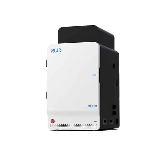
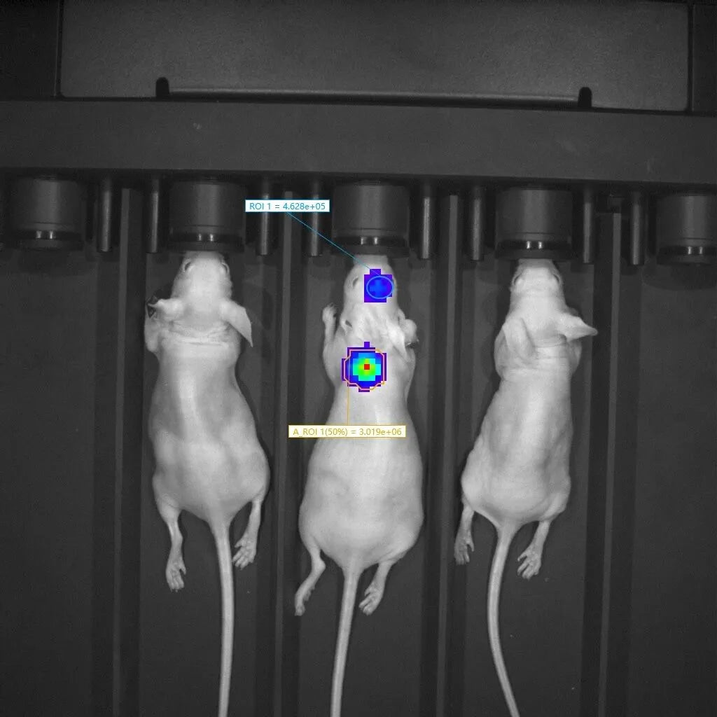
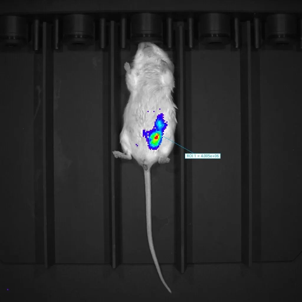
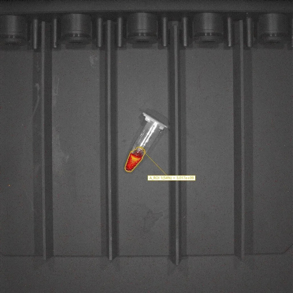
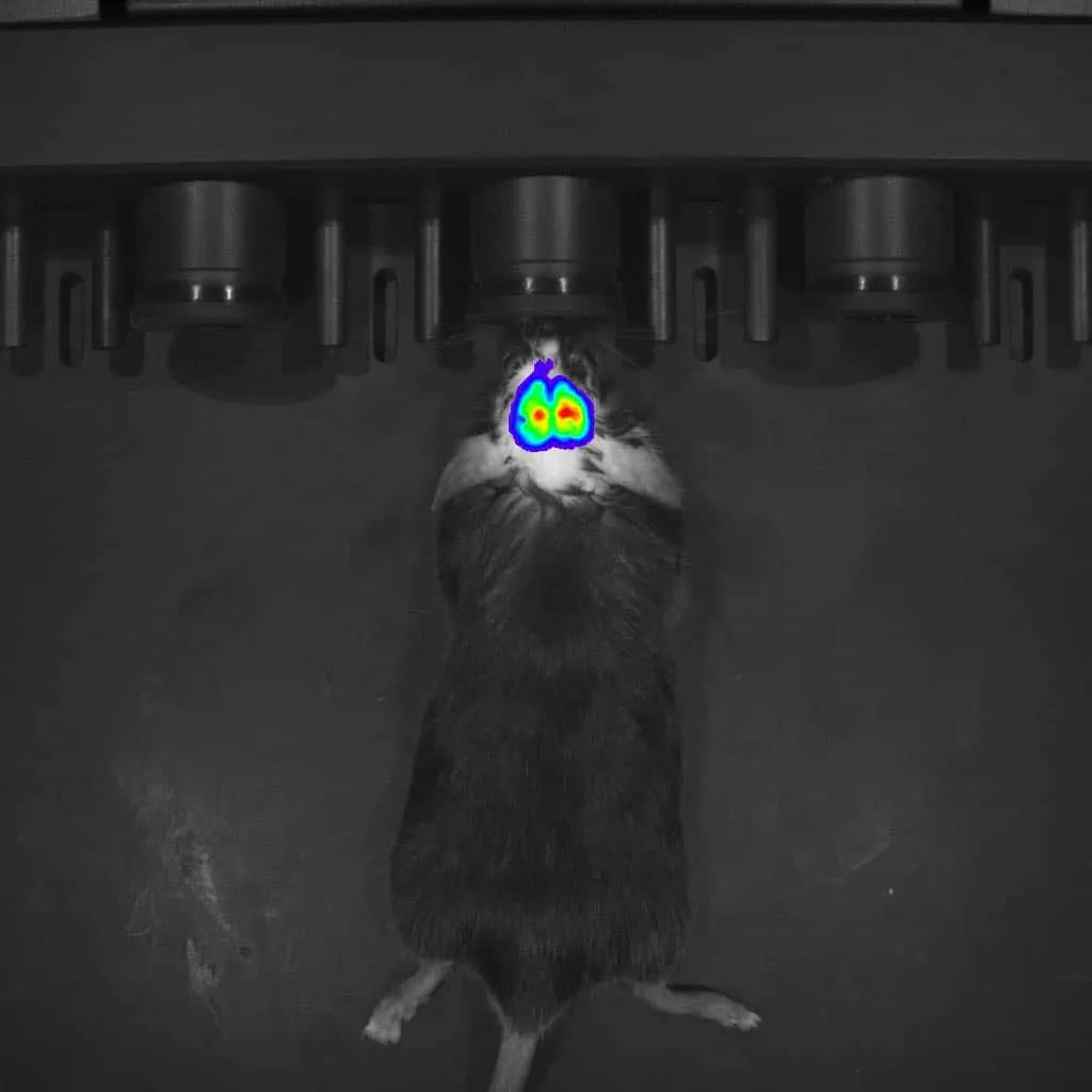
The MOIS Series (HTP / HT / HTX) is a high-sensitivity in vivo imaging system designed for small animal research. It integrates bioluminescence, fluorescence, and spectral imaging, enabling accurate, low-light detection across tumor monitoring, immune cell tracking, and pharmacokinetics. With up to -90°C CCD cooling and up to 26 narrowband filters, MOIS ensures high signal-to-noise performance, spectral precision, and quantitative reliability—making it a powerful platform for dynamic, multiscale biomedical studies.
The MOIS HT small animal in vivo imaging system is a non-invasive imaging system designed for real-time monitoring and visualization of biological processes in live small animals (such as mice and rats). Equipped with both bioluminescence and fluorescence imaging modes, this system allows researchers to observe molecular and cellular activities in living organisms, providing valuable insights into disease progression, treatment efficacy, and underlying biological mechanisms, all without the need for euthanizing the animals.
Small animal in vivo imaging technology is extensively used in preclinical research, particularly in areas such as oncology, cardiovascular research, neurology, drug development, as well as in gene therapy and regenerative medicine.
Ultra-High Sensitivity
Up to -90°C CCD cooling and ≥90% quantum efficiency enable exceptional low-light performance for bioluminescence and fluorescence imaging
Broad Spectral Compatibility
Excitation wavelengths from 400 to 900 nm support diverse fluorophores used in oncology, immunology, and stem cell research
Reliable Multimodal Imaging
Integrates bioluminescence, fluorescence, X-ray,and Cherenkov imaging in one system for versatile, in vivo experimental workflows.
Quantitative Accuracy
Calibration, spectral unmixing, and flat-field correction ensure reliable quantification across imaging sessions.
Scalable Field of View
Imaging range from 2.5 × 2.5 cm to 25 × 25 cm covers both localized and whole-body applications.
Data Consistency
Bias, background, and cosmic ray corrections enhance image uniformity and reproducibility.
Journal: Theranostics
Use the small animal in vivo imaging system to image the distribution of Mn₃O₄ nanozymes in ex vivo organs.

| Module | Parameters | MOIS HTP | MOIS HT | MOIS HTX |
| Imaging Modules | Fluorescence Imaging Module | √ | √ | √ |
| Bioluminescence Imaging Module | √ | √ | √ | |
| Spectral High-Resolution Imaging | √ | √ | √ | |
| X-Ray Module | × | × | √ | |
| Upconversion Fluorescence Imaging (UCFI) Module | Expandable & Upgradeable | |||
| Detector | Camera Type | Back-illuminated sensor, Scientific-grade CCD | ||
| Operating Temperature | -70°C | -90°C | -90°C | |
| Pixel Count | 1024×1024 | |||
| Aperture (F-Stop) | ≤ 0.95 | |||
| Minimum FOV (Field of View) | 2.5cm × 2.5cm (optional) | |||
| Maximum FOV | 25cm × 25cm | |||
| Quantum Efficiency | ≥90% (500–700nm) | |||
| Broadband Light Source | 150W Halogen-tungsten broadband | |||
| Laser System | Guide Laser | Real-time FOV center display for positioning | ||
| Filters Excitation Filters / Emission Filters |
19 / 7 | |||
| Software | Real-time Acquisition System | Supports real-time image acquisition with light prompts, time series module, communication connection, and bioluminescence/fluorescence multi-mode imaging switching. Synchronous acquisition module supported. | ||
| Offline Analysis Workstation | Support image analysis, time spectrum analysis module, need to be authorized by molecular software quantitative module | |||
| Environmental Control | Multi-Animal Imaging Chamber | 5 channels | ||
| Temperature Module | Temperature control range: 20-40°C, accuracy: ±0.1°C | |||
| Anesthesia System | Anesthesia gas control: isoflurane, concentration: 0.5%–5% adjustable | |||
| Model | Parameter | MOIS HTP | MOIS HT |
| -70°C Version | Dark Current | 0.0004 (typical) – 0.001 (max) | 0.000177 (typical) |
| Readout Noise | @100KHz: 3.0 e⁻ (typical), 5 e⁻ (max) @2MHz: 9 e⁻ (typical), 15 e⁻ (max) |
@50KHz: 2.9 e⁻ (typical) @1MHz: 6.6 e⁻ (typical) |
|
| Signal-to-Noise Ratio (SNR) | Slightly lower SNR, may affect high-precision imaging | Higher SNR, suitable for ultra-low noise high-resolution imaging | |
| Image Quality | Good for standard imaging, less effective for low light signals | Excellent low light capture, ideal for high-resolution and low noise imaging | |
| Application Scenarios | General small animal imaging experiments | High-precision imaging, low signal detection, long-term monitoring | |
| Stability | Higher dark current and noise may affect result consistency | Lower dark current and noise enhance image stability and reliability | |
| Research Suitability | Basic research and drug development | Preclinical research, oncology, drug tracking, and dynamic monitoring |
